Rhombencephalon: Structure And Functions
The brain is divided into parts to better understand its development and functions. One of these is the hindbrain, an area that originates from the caudal primary embryonic vesicle.
When we talk about the hindbrain , we refer to the hind brain. During its existence, this area gives rise to different sub-structures responsible for carrying out various essential functions for the organism.
In today’s article we will show you this structure, how the differentiation process takes place and the functions of this incredible drive center.
Differentiation of the hindbrain
To begin, we need to understand the origins of the hindbrain. To do this, it is important to understand what differentiation is. According to Bear, Connors and Paradiso, authors of the book Neuroscience. Exploring the brain, it is a process in which structures become more complex and specialize.
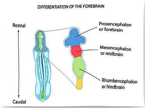
The first step in brain differentiation is the development of three compartments called primary vesicles of the neural tube that originate in the extreme rostral.
The most rostral part of the primary vesicles is the forebrain or forebrain; the vesicle located behind the forebrain is called the midbrain or middle brain, while the most caudal part of the vesicles is the hindbrain or posterior brain, which in turn connects with the caudal part of the neural tube.
The hindbrain, therefore, is formed during embryonic development, and does so through transverse segments called rhombomeres, compartments that allow the creation of cell groups that will develop in different ways and take on different functions. The hindbrain is divided into three essential structures:
- Cerebellum. It joins the brain stem with the bridge, and is a movement control center, essential for our body. It comes from the rostral area.
- Bridge. It is a part of the rostral hindbrain. It is located anterior to the brain and the fourth ventricle.
- Bulb or medulla oblongata. It is located caudal to the pons and cerebellum. It comes from the caudal area.
In the area of the vesicles, the rostral rhombencafalo has the shape of a tube. In the posterior part, the rhombic lip or tissue of the dorsolateral wall of the tube grows rostral and medial until it merges with the opposite side. The resulting fold grows to form the cerebellum. Finally, the ventral wall of the tube expands to form the bridge or protuberance.
On the other hand, in the differentiation of the caudal half of the posterior brain into the spinal bulb, changes occur, but less pronounced. On the one hand, the walls dilate and leave only the roof covered with non-neuronal ependymal cells. On the other hand, white matter systems are present on the entire ventral surface of each side of the medulla oblongata or spinal bulb.
Finally, the hole occupied by the cerebrospinal fluid transforms into the fourth ventricle, which will continue with the cerebral aqueduct of the midbrain.
Functions of the hindbrain
The hind brain performs several functions. Let’s see them:
- It is a key area for information, from the forebrain to the spinal cord and vice versa. For example, of white matter beams.
- Its neurons collaborate in the process of sensory information.
- Part of the hindbrain neurons contribute to the control of voluntary movement. In addition, they help regulate the autonomous system.
- The cerebellum, also called the small brain, regulates movement as if it were a control center. It also receives huge amounts of axons, coming from the spinal cord and the pons. On the other hand, the cerebellum is in charge of comparing the information that arrives and calculating the sequences of muscle contractions, essential for carrying out the movement.
- The spinal bulb has the task of carrying somatic information from the spinal cord to the thalamus. In addition, it controls the movements of the tongue and is associated with the sensory functions of touch and taste.
- The axons of the auditory nerves are responsible for carrying information from the ears to the cochlear nuclei of the bulb. The nuclei are in charge of projecting axons to different structures, including the roof of the midbrain.
Well, the stimuli that come from the spinal cord transmit information about the position of the body in space. In addition, the inputs of the bridge have the task of transmitting information from the cerebral cortex, as well as specifying the purpose of the movement.
Possible associated disorders
If brain development is inadequate, the hindbrain and its vital functions could be damaged. Let’s see what could happen:
- Injuries to the hindbrain can cause motor problems, such as uncoordinated and inaccurate movements, as in the case of ataxia.
- The damage could lead to deafness if, for example, a lesion occurs in the cochlear nuclei.
- Problems related to touch and taste.
- Dandy Walker and Arnold Chiari syndrome, i.e. resulting from the abnormal development of the hindbrain. The damage could cause vomiting, weakness, breathing and circulation problems.
- Rhombencephalitis, which is inflammation of the hindbrain caused by several factors.
As we have seen, the hindbrain is a fundamental part of our body. Through its motor, sensory and visceral functions, it regulates its functioning. If damaged or if not developed properly, it could lead to very serious consequences for our survival.