Neuroscience Research Tools
Neuroscience is a scientific discipline that studies the nervous system and the way in which the different elements that constitute it interact and define conduct. It is a complex field of study that spans from neuronal functioning to behavior and is therefore very broad. However, it is of great use to us in understanding how our conduct develops. This discipline uses the scientific method to obtain knowledge through a series of research tools.
These are useful in exploring brain anatomy and function. Each of them has some advantages and disadvantages that make them suitable in certain situations and not in others.
In this article we will briefly expose the most used research tools in neuroscience, namely EEG, MEG, CT, TEP and MRI.
Most used research tools in neuroscience
Electroencephalography (EEG)
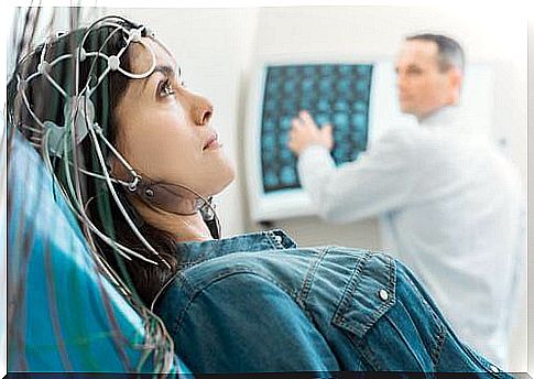
It is an instrument that measures how electricity flows through the cerebral cortex. When a neuron is activated, a passage of ions takes place through it that we can measure with a series of electrodes.
These electrodes are placed directly on the scalp together with a substance that facilitates the passage of current. It is thus possible to capture neuronal activity in the form of waves.
EEG is one of the neuroscience research tools with great temporal capacity. However, its space capacity is somewhat poor. It is useful for us to relate certain waves to certain processes, but if we wish to locate them we will have to use another tool.
It is used, for example, to analyze the different phases of sleep, because each being corresponds to a specific type of waves.
Magnetoencephalography (MEG)
It is very similar to the EEG, but it does not pick up changes in voltage, but rather the magnetic fields of neurons. Each electric current generates a magnetic field perpendicular to itself. This physical principle allows us to apply receptors on the scalp capable of measuring brain activity.
The structural anatomy of the cortex also prevents the magnetic field of some neurons from exiting the skull. This is useful for measuring the activity of certain brain areas without noise or interference.
Compared to the EEG, the MEG offers a worse temporal resolution, since the magnetic field detection has a longer delay. Nevertheless, it guarantees a better spatial resolution, since we can know the exact area in which these magnetic fields were generated.
Computed Axial Tomography (CT)
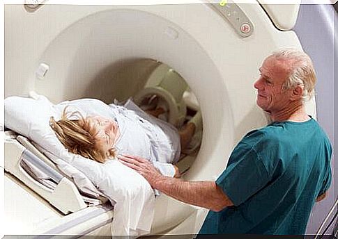
It is one of the most useful research tools for exploring the structural anatomy of the brain. It consists of passing a multitude of X-ray beams around the head from different angles. Then, through a computer program, all the images are merged to obtain a 3D image of the brain.
When they pass through the human body, part of the X-rays are absorbed by the affected structures. If we place a receptor on the other side, we can see a photograph of the X-ray residue. This will give us an image of the areas crossed in a gray scale.
CT is a very useful technique for viewing brain anatomy and has a very low cost, as well as being simple. Even so, it has certain drawbacks. The main one and perhaps the most serious is the invasiveness of the exam. Part of the radiation is absorbed by the brain, which forces it to be used limitedly to avoid any damage. In addition, nowadays there are techniques with better spatial and temporal resolution than CT, such as magnetic resonance.
Positron Emission Tomography (TEP)
The TEP allows to determine the level of metabolic activity of each area of the brain. A very useful tool for research, as it offers us a lot of information on where brain activity occurs.
For this purpose, the individual is injected with glucose bound to a radioactive molecule (2-deoxy-D-glucose). This substance will travel to the brain, where the positrons of the radioactive isotope will react with the electrons of surrounding atoms. In this way, they will mutually destroy each other by releasing light.
This light caused by the reaction of positrons can be picked up by a receptor, thus allowing to obtain an image of the areas of the brain that have consumed the most glucose.
This technique is often used in conjunction with CT to know exactly the structures in which glucose has been metabolized. The TEP has a high spatial resolution, but the temporal resolution is not the best, since it is necessary to wait for the substance to be consumed by the brain. In general, this process occurs after the cognitive event we wish to measure.
It is also one of the most invasive techniques among neuroscientific research tools. In fact, it requires the introduction of radiation directly into the brain, with the consequent danger for its structures. In light of this, it is used only when strictly necessary.
Magnetic resonance imaging (MRI) and functional magnetic resonance imaging (MRI)
Along with CT, MRI is one of the most used techniques in neuroscience and medicine. MRI takes advantage of the fact that the atoms of certain substances in the human body react when an electromagnetic wave passes through them.
A large magnet is used to orient the axis of all the hydrogen atoms in the brain in one direction. When the electromagnetic pulse ceases, all these atoms return to their place, returning an energy signal that we can detect.
The RMF is a variant of the first that allows us to measure activity and brain structure in real time while the subject performs an activity with low temporal latency. Among the neuroscientific research tools it is perhaps the one that offers the best spatial and temporal results.
Its invasiveness is totally nil, since magnetic fields below a certain power do not damage the brain structure. Its problem lies, however, in its very high costs, both of the staff and of its maintenance. Getting an MRI machine costs around five million euros, which is why not all hospitals can afford one.
In this article you have been able to discover some of the research tools currently used in neuroscience. This field of study is still in its early stages, but thanks to these techniques we are increasingly getting to know our brain and how it works.